
Rosalind Franklin and the Hidden DNA Photograph
A patient scientist, a mysterious X-shaped pattern and a photograph that helped reveal the structure of life. This is the story of Rosalind Franklin, Photograph 51 and the scientific evidence that changed biology.
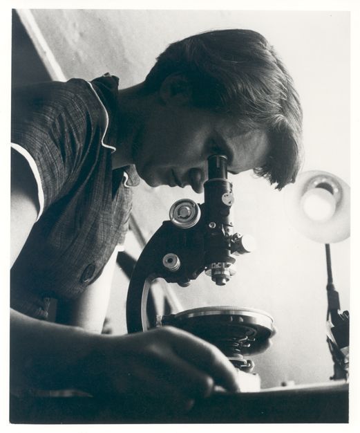 Rosalind Franklin, 1955
Image: MRC Laboratory of Molecular Biology, from the personal collection of Jenifer Glynn. Licensed under CC BY-SA 4.0 via Wikimedia Commons.
Four facts before the mystery begins
Franklin was far more than a single photograph. She was a skilled physical chemist and X-ray crystallographer whose research crossed several fields.
Born in London
Rosalind Elsie Franklin was born on 25 July 1920 and showed an early strength in science and logical problem-solving.
Photograph 51
Franklin and her research student Raymond Gosling produced the famous X-ray diffraction image of hydrated B-form DNA.
Hours of exposure
The DNA fibres were exposed to X-rays for roughly 62 hours before the exceptionally clear pattern was developed.
A short but powerful career
Franklin died in 1958 at the age of 37, after making major contributions to research on DNA, coal, graphite and viruses.
A scientific mystery hidden in scattered X-rays
Move through the chapters to see how careful experiments, difficult working relationships and shared scientific evidence shaped the discovery of DNA's structure.
A young mind that wanted evidence
Rosalind Franklin grew up in London in a family that valued education and public service. At school, she became known for her ability in mathematics and science.
She later studied physical chemistry at Newnham College, Cambridge. Science was still a difficult profession for women, but Franklin developed the disciplined thinking that would define her career: measure carefully, repeat experiments and do not accept a model until the evidence supports it.
Coal, graphite and a laboratory in Paris
During the Second World War, Franklin studied coal and related carbon materials. Her work helped explain why different kinds of coal behaved differently when heated and how their microscopic structures affected their industrial use.
She then moved to Paris, where she became highly skilled in X-ray crystallography. In this technique, X-rays are directed at a carefully prepared sample. The atoms scatter the rays, producing a pattern that scientists can use to calculate the arrangement of matter inside the sample.
It was not an ordinary photograph. It was a mathematical message written in spots, shadows and distances.
The difficult DNA laboratory
Franklin joined the biophysics research unit at King's College London in 1951. She was asked to investigate DNA fibres using X-ray diffraction.
The working arrangement was troubled from the beginning. Responsibilities were not communicated clearly, and Franklin and Maurice Wilkins did not agree on how their roles were organised. Their scientific styles and personalities also differed.
Despite the tension, Franklin improved the laboratory equipment, controlled the moisture around the DNA samples and separated two different forms of DNA. The drier form was called A-DNA, while the wetter and more clearly helical form was called B-DNA.
The appearance of Photograph 51
Franklin and her PhD student Raymond Gosling prepared a thin fibre containing many similarly aligned DNA molecules. They exposed it to X-rays for about 62 hours.
When the film was developed, it revealed a striking X-shaped arrangement of spots. The image was numbered 51 in the laboratory series, which is why it became known as Photograph 51 or Photo 51.
The X shape strongly suggested a helix. The spacing of the spots also contained information about the dimensions of DNA and the repeating arrangement inside the molecule.
The photograph was shown without her knowledge
Maurice Wilkins showed a copy of Photograph 51 to James Watson. Franklin did not know that the image had been shown to him.
Watson immediately recognised how clearly the X-shaped pattern supported a helical structure. Other measurements from Franklin's work also reached Watson and Francis Crick through a research report.
Historians continue to examine the scientific culture, assumptions and communication failures surrounding these events. What is clear is that Franklin's experimental results became important to the successful model built by Watson and Crick.
Three papers and one double helix
Watson and Crick completed their double-helix model. Their paper appeared in the journal Nature on 25 April 1953.
It was followed in the same issue by papers from the King's College researchers, including a paper by Franklin and Gosling. Readers could see that the proposed model agreed with the X-ray evidence.
Franklin was also analysing the evidence independently. A draft she prepared in early 1953 concluded that a helical structure was highly probable and identified important features such as the position of the phosphate groups and the number of bases in a complete turn.
Her contribution was essential, but the full significance of her role was not widely understood by the public until much later.
A career that continued beyond DNA
Franklin moved to Birkbeck College, where she led important research on viruses. Her group studied the tobacco mosaic virus and contributed to research on the poliovirus.
She worked with scientists including Aaron Klug, who later received the Nobel Prize in Chemistry for his own structural research. Franklin's virus work helped establish methods that continued to influence molecular biology.
She died from ovarian cancer on 16 April 1958. Four years later, Watson, Crick and Wilkins received the 1962 Nobel Prize in Physiology or Medicine for discoveries concerning the molecular structure of nucleic acids.
Franklin was not among the recipients. Today, museums, universities, scientific awards, laboratories and educational programmes carry her name and recognise the depth of her work.
Develop a diffraction-inspired image
Press the button to reveal an educational reconstruction of an X-shaped diffraction pattern. Then investigate the scientific clues.
The undeveloped plate
The visual below is an original educational illustration inspired by diffraction patterns. It is not a reproduction of Photograph 51.
Educational visualisation created with CSS for Speed Up Science.
Read the scientific clues
Select each clue to understand what a diffraction pattern can reveal.
One discovery, several kinds of evidence
The structure of DNA emerged from combined experimental results and model-building rather than from one person or one photograph alone.
Precise experiments
Franklin controlled humidity, improved the X-ray equipment and produced highly detailed diffraction patterns of DNA fibres.
Chemical information
Scientists already knew that DNA contained sugars, phosphates and four nitrogenous bases. The proposed structure had to follow the rules of chemistry.
Base ratios
Erwin Chargaff had shown that the amount of adenine was close to thymine, while cytosine was close to guanine.
Mathematical measurements
The locations of diffraction spots helped scientists estimate the width of DNA and the repeating distance along its helix.
Model-building
Watson and Crick tested physical arrangements until they produced a double helix that agreed with the known chemical and experimental evidence.
Scientific publishing
The 1953 Nature papers presented the proposed model together with experimental work from the King's College research groups.
What the popular version often leaves out
Open each statement to separate a dramatic myth from the more accurate historical picture.
Myth: Franklin did not understand her own photograph.
Myth: Photograph 51 solved the entire structure by itself.
Myth: Franklin was working as an assistant.
Myth: Franklin and the other DNA researchers were one harmonious team.
Myth: Her entire scientific career was about DNA.
The scientist behind much more than Photo 51
Research contributions
Franklin repeatedly used physical evidence to answer difficult questions about materials and biological structures.
- Fundamental research on coal and graphite
- Expert use of X-ray crystallography
- Identification of distinct A and B forms of DNA
- Detailed measurements of DNA's structure
- Research on tobacco mosaic virus
- Contributions to structural studies of poliovirus
Why her story matters
Scientific discoveries depend on evidence, but recognition also depends on communication, professional culture and the way history is recorded.
Franklin's story encourages laboratories to give clear credit, define responsibilities, share data ethically and recognise experimental work as strongly as celebrated theories and models.
It also reminds students that careful measurement can be as creative and revolutionary as a dramatic flash of inspiration.
The lesson hidden inside the photograph
A scientific image does not speak for itself. Someone must design the experiment, prepare the sample, control the conditions, calculate the measurements and understand what the pattern means. Photograph 51 is powerful because it represents all of that invisible work.
Can you solve the DNA story quiz?
Answer all five questions and press the button to calculate your score.
1. Who produced Photograph 51?
2. What did the X-shaped pattern strongly suggest?
3. Which form of DNA appeared in Photograph 51?
4. Which statement about Franklin's career is accurate?
5. Did Photograph 51 solve DNA's structure completely by itself?
Questions about Rosalind Franklin and Photo 51
What was Photograph 51?
Who took Photograph 51?
Why was the X shape important?
Was Photograph 51 shown without Franklin's knowledge?
Did Rosalind Franklin understand that DNA was helical?
Why did Rosalind Franklin not receive the 1962 Nobel Prize?
What else did Rosalind Franklin study?
Learn science beyond the textbook
Follow Speed Up Science on Instagram for student-friendly science stories, visual explanations, activities and learning challenges.
Follow @speedupscience_





0 Comments